Ключевые слова
моноклональная гаммапатия неопределенного значения
болезнь холодовых агглютининов
лимфоплазмоцитарная лимфома
AL-амилоидоз
Как цитировать
Статистика
PDF_2024-17-2_94-108 загрузок: 840
Ключевые слова
Аннотация
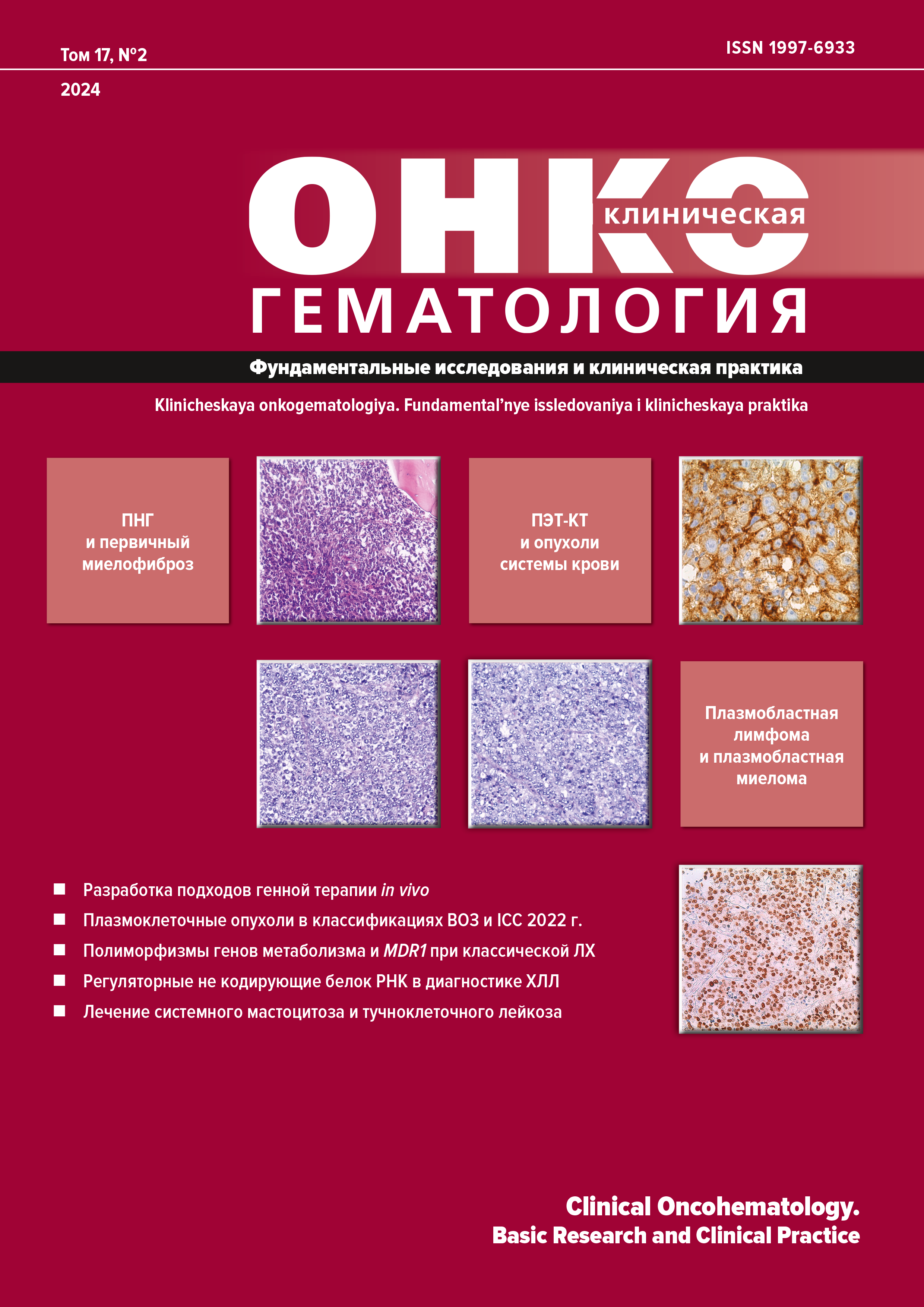
В 2022 г. гематологическое сообщество столкнулось с весьма нетривиальным событием одновременной публикации двух конкурирующих классификаций опухолей кроветворной и лимфоидной тканей, подготовленных разными группами ведущих международных экспертов. На протяжении последних 20 лет общепризнанным стандартом диагностики служило несколько последовательных ревизий классификаций гематологических новообразований, опубликованных Всемирной организацией здравоохранения (ВОЗ) в 2001, 2008 и 2016 гг. Со времени издания 4-го пересмотра классификации ВОЗ (WHO-HAEM4) были накоплены новые клинико-патологические, биологические и молекулярные знания в данной области, что способствовало уточнению диагностических критериев ряда заболеваний, появлению новых терминов и утверждению понятий, ранее определяемых как требующие дальнейшего уточнения. Как результат была подготовлена очередная, уже 5-я по счету, редакция классификации ВОЗ опухолей кроветворной и лимфоидной тканей (WHO-HAEM5), опубликованная в виде предварительной статьи в журнале «Leukemia». При этом важно отметить, что окончательная версия «Синей книги ВОЗ» в 2023 г. так и не увидела свет, поэтому еще возможно внесение в нее отдельных дополнений. Кроме того, в том же 2022 г. в журнале «Blood» выходит статья «Международная консенсусная классификация зрелых лимфоидных опухолей», сокращенно — ICC. Авторский состав двух классификаций практически не пересекается. В настоящей обзорной статье в сравнительном аспекте рассматриваются две классификации с точки зрения новых диагностических критериев и верификации конкретных клинико-морфологических категорий. Преимущественно обзор сосредоточен на описании плазмоклеточных опухолей и родственных им В-клеточных лимфопролиферативных заболеваний, протекающих с секрецией моноклонального иммуноглобулина.
Библиографические ссылки
- Alaggio R, Amador C, Anagnostopoulos I, et al. The 5th edition of the World Health Organization Classification of Haematolymphoid Tumours: Lymphoid Neoplasms. Leukemia. 2022;36(7):1720–48. doi: 10.1038/s41375-022-01620-2.
- Campo E, Jaffe ES, Cook JR, et al. The International Consensus Classification of Mature Lymphoid Neoplasms: a report from the Clinical Advisory Committee. Blood. 2022;140(11):1229–53. doi: 10.1182/blood.2022015851.
- Kyle RA, Ansell SM, Kapoor P. Prognostic factors and indications for treatment of Waldenstrom’s Macroglobulinemia. Best Pract Res Clin Haematol. 2016;29(2):179–86. doi: 10.1016/j.beha.2016.08.014.
- Khwaja J, D’Sa S, Minnema MC, et al. IgM monoclonal gammopathies of clinical significance: diagnosis and management. Haematologica. 2022;107(9):2037–50. doi: 10.3324/haematol.2022.280953.
- Annibali O, Petrucci MT, Del Bianco P, et al. IgM multiple myeloma: report of four cases and review of the literature. Leuk Lymphoma. 2006;47(8):1565–9. doi: 10.1080/10428190600604450.
- Kyle RA, Anderson KC. A tribute to Jan Gosta Waldenstrom. Blood. 1997;89(12):4245–7.
- Owen RG, Treon SP, Al-Katib A, et al. Clinicopathological definition of Waldenstrom’s macroglobulinemia: consensus panel recommendations from the Second International Workshop on Waldenstrom’s Macroglobulinemia. Semin Oncol. 2003;30(2):110–5. doi: 10.1053/sonc.2003.50082.
- Fend F, Dogan A, Cook JR. Plasma cell neoplasms and related entities-evolution in diagnosis and classification. Virchows Arch. 2023;482(1):163–77. doi: 10.1007/s00428-022-03431-3.
- Garcia-Abellas P, Ferrer Gomez A, Bueno Sacristan D, et al. Lymphoplasmacytic lymphoma and marginal zone lymphoma involving bone marrow: A diagnostic dilemma. Useful clinicopathological features to accurate the diagnosis. EJHaem. 2022;3(4):1181–7. doi: 10.1002/jha2.573.
- Kivrak H, Yuksel S, Ates C, et al. Relevance of Additional Immunohistochemical Markers in the Differential Diagnosis of Small B-Cell Lymphomas: A Case-Control Study. Turk J Haematol. 2022;39(3):178–87. doi: 10.4274/tjh.galenos.2021.2021.0349.
- Gertz MA. Waldenstrom macroglobulinemia: 2023 update on diagnosis, risk stratification, and management. Am J Hematol. 2023;98(2):348–58. doi: 10.1002/ajh.26796.
- Treon SP, Xu L, Guerrera ML, et al. Genomic Landscape of Waldenstrom Macroglobulinemia and Its Impact on Treatment Strategies. J Clin Oncol. 2020;38(11):1198–208. doi: 10.1200/JCO.19.02314.
- Deguine J, Barton GM. MyD88: a central player in innate immune signaling. F1000Prime Rep. 2014;6:97. doi: 10.12703/P6-97.
- Awata-Shiraiwa M, Yokohama A, Kanai Y, et al. Waldenstrom macroglobulinemia and non-IgM-type lymphoplasmacytic lymphoma are genetically similar. Acta Haematol. 2023;146(5):384–90. doi: 10.1159/000530100.
- Treon SP, Xu L, Hunter Z. MYD88 Mutations and Response to Ibrutinib in Waldenstrom’s Macroglobulinemia. N Engl J Med. 2015;373(6):584–6. doi: 10.1056/NEJMc1506192.
- Kaiser LM, Hunter ZR, Treon SP, Buske C. CXCR4 in Waldenstrom’s Macroglobulinema: chances and challenges. Leukemia. 2021;35(2):333–45. doi: 10.1038/s41375-020-01102-3.
- Alcoceba M, Garcia-Alvarez M, Medina A, et al. MYD88 Mutations: Transforming the Landscape of IgM Monoclonal Gammopathies. Int J Mol Sci. 2022;23(10):5570. doi: 10.3390/ijms23105570.
- Garcia-Sanz R, Dogliotti I, Zaccaria GM, et al. 6q deletion in Waldenstrom macroglobulinaemia negatively affects time to transformation and survival. Br J Haematol. 2021;192(5):843–52. doi: 10.1111/bjh.17028.
- Krzisch D, Guedes N, Boccon-Gibod C, et al. Cytogenetic and molecular abnormalities in Waldenstrom’s macroglobulinemia patients: Correlations and prognostic impact. Am J Hematol. 2021;96(12):1569–79. doi: 10.1002/ajh.26339.
- Nguyen-Khac F, Lambert J, Chapiro E, et al. Chromosomal aberrations and their prognostic value in a series of 174 untreated patients with Waldenstrom’s macroglobulinemia. Haematologica. 2013;98(4):649–54. doi: 10.3324/haematol.2012.070458.
- Hill QA, Stamps R, Massey E, et al. Guidelines on the management of drug-induced immune and secondary autoimmune, haemolytic anaemia. Br J Haematol. 2017;177(2):208–20. doi: 10.1111/bjh.14654.
- Jager U, Barcellini W, Broome CM, et al. Diagnosis and treatment of autoimmune hemolytic anemia in adults: Recommendations from the First International Consensus Meeting. Blood Rev. 2020;41:100648. doi: 10.1016/j.blre.2019.100648.
- Roelcke D. Cold agglutination. Antibodies and antigens. Clin Immunol Immunopathol. 1974;2(2):266–80. doi: 10.1016/0090-1229(74)90044-0.
- Berentsen S, Ulvestad E, Langholm R, et al. Primary chronic cold agglutinin disease: a population based clinical study of 86 patients. Haematologica. 2006;91(4):460–6.
- Berentsen S. How I treat cold agglutinin disease. Blood. 2021;137(10):1295–303. doi: 10.1182/blood.2019003809.
- Randen U, Troen G, Tierens A, al. Primary cold agglutinin-associated lymphoproliferative disease: a B-cell lymphoma of the bone marrow distinct from lymphoplasmacytic lymphoma. Haematologica. 2014;99(3):497–504. doi: 10.3324/haematol.2013.091702.
- Malecka A, Troen G, Tierens A, et al. Immunoglobulin heavy and light chain gene features are correlated with primary cold agglutinin disease onset and activity. Haematologica. 2016;101(9):e361–e364. doi: 10.3324/haematol.2016.146126.
- Berentsen S. New Insights in the Pathogenesis and Therapy of Cold Agglutinin-Mediated Autoimmune Hemolytic Anemia. Front Immunol. 2020;11:590. doi: 10.3389/fimmu.2020.00590.
- Malecka A, Delabie J, Ostlie I, et al. Cold agglutinin-associated B-cell lymphoproliferative disease shows highly recurrent gains of chromosome 3 and 12 or 18. Blood Adv. 2020;4(6):993–6. doi: 10.1182/bloodadvances.2020001608.
- Malecka A, Troen G, Tierens A, et al. Frequent somatic mutations of KMT2D (MLL2) and CARD11 genes in primary cold agglutinin disease. Br J Haematol. 2018;183(5):838–42. doi: 10.1111/bjh.15063.
- Boyle EM, Deshpande S, Tytarenko R, et al. The molecular make up of smoldering myeloma highlights the evolutionary pathways leading to multiple myeloma. Nat Commun. 2021;12(293):2. doi: 10.1038/s41467-41020-20524-41462.
- Rajkumar SV, Dimopoulos MA, Palumbo A, et al. International Myeloma Working Group updated criteria for the diagnosis of multiple myeloma. Lancet Oncol. 2014;15(12):e538–e548. doi: 10.1016/S1470-2045(14)70442-5.
- Kyle RA, Larson DR, Therneau TM, et al. Long-Term Follow-up of Monoclonal Gammopathy of Undetermined Significance. N Engl J Med. 2018;378(3):241–9. doi: 10.1056/NEJMoa1709974.
- Al Hamed R, Bazarbachi AH, Bazarbachi A, et al. Comprehensive Review of AL amyloidosis: some practical recommendations. Blood Cancer J. 2021;11(5):97. doi: 10.1038/s41408-021-00486-4.
- Leung N, Bridoux F, Batuman V, et al. The evaluation of monoclonal gammopathy of renal significance: a consensus report of the International Kidney and Monoclonal Gammopathy Research Group. Nat Rev Nephrol. 2019;15(1):45–59. doi: 10.1038/s41581-018-0077-4.
- Смирнов А.В., Афанасьев Б.В., Поддубная И.В. и др. Моноклональная гаммапатия ренального значения: консенсус гематологов и нефрологов России по введению нозологии, диагностике и обоснованности клон-ориентированной терапии. Нефрология. 2019;23(6):9–28. doi: 10.36485/1561-6274-2019-236-9-28. [Smirnov AV, Afanasyev BV, Poddubnaya IV, et al. Monoclonal gammopathy of renal significance: Consensus of hematologists and nephrologists of Russia on the establishment of nosology, diagnostic approach and rationale for clone specific treatment. Nephrology (Saint-Petersburg). 2019;23(6):9–28. doi: 10.36485/1561-6274-2019-236-9-28. (In Russ)]
- Amaador K, Peeters H, Minnema MC, et al. Monoclonal gammopathy of renal significance (MGRS) histopathologic classification, diagnostic workup, and therapeutic options. Neth J Med. 2019;77(7):243–54.
- Leung N, Bridoux F, Hutchison CA, et al. Monoclonal gammopathy of renal significance: when MGUS is no longer undetermined or insignificant. Blood. 2012;120(22):4292–5. doi: 10.1182/blood-2012-07-445304.
- Rajkumar SV, Kumar S, Lonial S, Mateos MV. Smoldering multiple myeloma current treatment algorithms. Blood Cancer J. 2022;12(9):129. doi: 10.1038/s41408-022-00719-0.
- Ravindran A, Bartley AC, Holton SJ, et al. Prevalence, incidence and survival of smoldering multiple myeloma in the United States. Blood Cancer J. 2016;6(10):e486. doi: 10.1038/bcj.2016.100.
- Kyle RA, Remstein ED, Therneau TM, et al. Clinical course and prognosis of smoldering (asymptomatic) multiple myeloma. N Engl J Med. 2007;356(25):2582–90. doi: 10.1056/NEJMoa070389.
- Lakshman A, Rajkumar SV, Buadi FK, et al. Risk stratification of smoldering multiple myeloma incorporating revised IMWG diagnostic criteria. Blood Cancer J. 2018;8(6):59. doi: 10.1038/s41408-018-0077-4.
- Vaxman I, Gertz MA. How I approach smoldering multiple myeloma. Blood. 2022;140(8):828–38. doi: 10.1182/blood.2021011670.
- Moreau P, San Miguel J, Sonneveld P, et al. Multiple myeloma: ESMO Clinical Practice Guidelines for diagnosis, treatment and follow-up. Ann Oncol. 2017;28(Suppl_4):iv52–iv61. doi: 10.1093/annonc/mdx096.
- Kind S, Merenkow C, Buscheck F, et al. Prevalence of Syndecan-1 (CD138) Expression in Different Kinds of Human Tumors and Normal Tissues. Dis Markers. 2019;2019:4928315. doi: 10.1155/2019/4928315.
- Flores-Montero J, de Tute R, Paiva B, et al. Immunophenotype of normal vs. myeloma plasma cells: Toward antibody panel specifications for MRD detection in multiple myeloma. Cytometry B Clin Cytom. 2016;90(1):61–72. doi: 10.1002/cyto.b.21265.
- An G, Xu Y, Shi L, et al. t(11;14) multiple myeloma: a subtype associated with distinct immunological features, immunophenotypic characteristics but divergent outcome. Leuk Res. 2013;37(10):1251–7. doi: 10.1016/j.leukres.2013.06.020.
- Fonseca R, Bergsagel PL, Drach J, et al. International Myeloma Working Group molecular classification of multiple myeloma: spotlight review. Leukemia. 2009;23(12):2210–21. doi: 10.1038/leu.2009.174.
- Tian E, Sawyer JR, Heuck CJ, et al. In multiple myeloma, 14q32 translocations are nonrandom chromosomal fusions driving high expression levels of the respective partner genes. Genes Chromosomes Cancer. 2014;53(7):549–57. doi: 10.1002/gcc.22165.
- Bal S, Kumar SK, Fonseca R, et al. Multiple myeloma with t(11;14): unique biology and evolving landscape. Am J Cancer Res. 2022;12(7):2950–65.
- Prideaux SM, Conway O’Brien E, Chevassut TJ. The genetic architecture of multiple myeloma. Adv Hematol. 2014;2014:864058. doi: 10.1155/2014/864058.
- Fonseca R, Barlogie B, Bataille R, et al. Genetics and cytogenetics of multiple myeloma: a workshop report. Cancer Res. 2004;64(4):1546–58. doi: 10.1158/0008-5472.can-03-2876.
- Abdallah N, Rajkumar SV, Greipp P, et al. Cytogenetic abnormalities in multiple myeloma: association with disease characteristics and treatment response. Blood Cancer J. 2020;10(8):82. doi: 10.1038/s41408-020-00348-5.
- D’Agostino M, Cairns DA, Lahuerta JJ, et al. Second Revision of the International Staging System (R2-ISS) for Overall Survival in Multiple Myeloma: A European Myeloma Network (EMN) Report Within the HARMONY Project. J Clin Oncol. 2022;40(29):3406–18. doi: 10.1200/JCO.21.02614.
- Войцеховский В.В., Григоренко А.А., Есенина Т.В. и др. Особенности диагностики и лечения различных вариантов плазмоцитомы. Бюллетень физиологии и патологии дыхания. 2023;88:105–19. doi: 10.36604/1998-5029-2023-88-105-119. [Voytsekhovskiy VV, Grigorenko AA, Esenina TV, et al. Features of diagnostics and treatment of various plasmacytoma options. Bulletin Physiology and Pathology of Respiration. 2023;88:105–19. doi: 10.36604/1998-5029-2023-88-105-119. (In Russ)]
- Caers J, Paiva B, Zamagni E, et al. Diagnosis, treatment, and response assessment in solitary plasmacytoma: updated recommendations from a European Expert Panel. J Hematol Oncol. 2018;11(1):10. doi: 10.1186/s13045-017-0549-1.
- Nahi H, Genell A, Walinder G, et al. Incidence, characteristics, and outcome of solitary plasmacytoma and plasma cell leukemia. Population-based data from the Swedish Myeloma Register. Eur J Haematol. 2017;99(3):216–22. doi: 10.1111/ejh.12907.
- Swerdlow SH, Campo E, Pileri SA, et al. The 2016 revision of the World Health Organization classification of lymphoid neoplasms. Blood. 2016;127(20):2375–90. doi: 10.1182/blood-2016-01-643569.
- De Waal EG, Leene M, Veeger N, et al. Progression of a solitary plasmacytoma to multiple myeloma. A population-based registry of the northern Netherlands. Br J Haematol. 2016;175(4):661–7. doi: 10.1111/bjh.14291.
- Kremer M, Ott G, Nathrath M, et al. Primary extramedullary plasmacytoma and multiple myeloma: phenotypic differences revealed by immunohistochemical analysis. J Pathol. 2005;205(1):92–101. doi: 10.1002/path.1680.
- Firsova MV, Mendeleeva LP, Kovrigina AM, et al. Plasmacytoma in patients with multiple myeloma: morphology and immunohistochemistry. BMC Cancer. 2020;20(1):346. doi: 10.1186/s12885-020-06870-w.
- Boll M, Parkins E, O’Connor SJ, et al. Extramedullary plasmacytoma are characterized by a ‘myeloma-like’ immunophenotype and genotype and occult bone marrow involvement. Br J Haematol. 2010;151(5):525–7. doi: 10.1111/j.1365-2141.2010.08386.x.
- Mori H, Fukatsu M, Ohkawara H, et al. Heterogeneity in the diagnosis of plasmablastic lymphoma, plasmablastic myeloma, and plasmablastic neoplasm: a scoping review. Int J Hematol. 2021;114(6):639–52. doi: 10.1007/s12185-021-03211-w.
- Gowin K, Skerget S, Keats JJ, et al. Plasma cell leukemia: A review of the molecular classification, diagnosis, and evidenced-based treatment. Leuk Res. 2021;111:106687. doi: 10.1016/j.leukres.2021.106687.
- Sant M, Allemani C, Tereanu C, et al. Incidence of hematologic malignancies in Europe by morphologic subtype: results of the HAEMACARE project. Blood. 2010;116(19):3724–34. doi: 10.1182/blood-2010-05-282632.
- Gonsalves WI, Rajkumar SV, Go RS, et al. Trends in survival of patients with primary plasma cell leukemia: a population-based analysis. Blood. 2014;124(6):907–12. doi: 10.1182/blood-2014-03-565051.
- Kyle RA, Maldonado JE, Bayrd ED. Plasma cell leukemia. Report on 17 cases. Arch Intern Med. 1974;133(5):813–8. doi: 10.1001/archinte.133.5.813.
- Granell M, Calvo X, Garcia-Guinon A, et al. Prognostic impact of circulating plasma cells in patients with multiple myeloma: implications for plasma cell leukemia definition. Haematologica. 2017;102(6):1099–104. doi: 10.3324/haematol.2016.158303.
- Ravi P, Kumar SK, Roeker L, et al. Revised diagnostic criteria for plasma cell leukemia: results of a Mayo Clinic study with comparison of outcomes to multiple myeloma. Blood Cancer J. 2018;8(12):116. doi: 10.1038/s41408-018-0140-1.
- Fernandez de Larrea C, Kyle R, Rosinol L, et al. Primary plasma cell leukemia: consensus definition by the International Myeloma Working Group according to peripheral blood plasma cell percentage. Blood Cancer J. 2021;11(12):192. doi: 10.1038/s41408-021-00587-0.
- Bardwick PA, Zvaifler NJ, Gill GN, et al. Plasma cell dyscrasia with polyneuropathy, organomegaly, endocrinopathy, M protein, and skin changes: the POEMS syndrome. Report on two cases and a review of the literature. Medicine (Baltimore). 1980;59(4):311–22. doi: 10.1097/00005792-198007000-00006.
- Dispenzieri A. POEMS syndrome: 2021 Update on diagnosis, risk-stratification, and management. Am J Hematol. 2021;96(7):872–88. doi: 10.1002/ajh.26240.
- Пирадов М.А., Супонева Н.А., Гинзберг М.А. и др. POEMS-синдром: обзор литературы и описание клинических наблюдений. Журнал неврологии и психиатрии им. С.С. Корсакова. 2014;114(4):4–10. [Piradov MA, Suponeva NA, Ginzberg MA, et al. POEMS-syndrome: a literature review and case reports. Zhurnal Nevrologii i Psikhiatrii imeni S.S. Korsakova. 2014;114(4):4–10. (In Russ)]
- Михайлов А.М., Бессмельцев С.С., Пожарисский К.М. и др. Болезнь Каслмана и POEMS-синдром. Клиническая онкогематология. 2010;3(3):259–69. [Mihailov AM, Bessmeltcev SS, Pojarisskiy KM, et al. Castleman’s disease and POEMS-syndrome. Klinicheskaya onkogematologiya. 2010;3(3):259–69. (In Russ)]
- Sykes DB, O’Connell C, Schroyens W. The TEMPI syndrome. Blood. 2020;135(15):1199–203. doi: 10.1182/blood.2019004216.
- Lipsker D. Monoclonal gammopathy of cutaneous significance: review of a relevant concept. J Eur Acad Dermatol Venereol. 2017;31(1):45–52. doi: 10.1111/jdv.13847.
- Rosado FG, Oliveira JL, Sohani AR, et al. Bone marrow findings of the newly described TEMPI syndrome: when erythrocytosis and plasma cell dyscrasia coexist. Mod Pathol. 2015;28(3):367–72. doi: 10.1038/modpathol.2014.117.
- Lipsker D, Rondeau M, Massard G, Grosshans E. The AESOP (adenopathy and extensive skin patch overlying a plasmacytoma) syndrome: report of 4 cases of a new syndrome revealing POEMS (polyneuropathy, organomegaly, endocrinopathy, monoclonal protein, and skin changes) syndrome at a curable stage. Medicine (Baltimore). 2003;82(1):51–9. doi: 10.1097/00005792-200301000-00005.
- Lenormand C, Marzolf G, Lipsker D. AESOP syndrome: a potential life-saving and early clue to the diagnosis of POEMS syndrome. Clin Dermatol. 2021;39(2):215–9. doi: 10.1016/j.clindermatol.2020.10.002.
- Dagrosa AT, Cowdrey MCE, LeBlanc RE, et al. Adenopathy and extensive skin patch overlying a plasmacytoma with unusual histologic findings in a patient with polyneuropathy, organomegaly, endocrinopathy, monoclonal protein and skin changes syndrome and Castleman disease. J Cutan Pathol. 2019;46(10):784–9. doi: 10.1111/cup.13514.
- Быкова В.П., Дайхес Н.А., Платонова Г.А. и др. Опухолевидный амилоидоз верхних дыхательных путей. Архив патологии. 2019;81(5):74–9. doi: 10.17116/patol20198105174. [Bykova VP, Daikhes NA, Platonova GA, et al. Tumor-like amyloidosis of the upper respiratory tract. Arkhiv patologii. 2019;81(5):74–9. doi: 10.17116/patol20198105174. (In Russ)]
- Basset M, Hummedah K, Kimmich C, et al. Localized immunoglobulin light chain amyloidosis: Novel insights including prognostic factors for local progression. Am J Hematol. 2020;95(10):1158–69. doi: 10.1002/ajh.25915.
- Охота В.К., Рыжко В.В., Ковригина А.М. и др. Болезнь тяжелых цепей-μ в сочетании с системным амилоидозом и неамилоидными депозитами. Трудности диагностики и терапии. Гематология и трансфузиология. 2020;65(2):190–207. doi: 10.35754/0234-5730-2020-65-2-190-207. [Okhota VK, Ryzhko VV, Kovrigina AM, et al. μ-Heavy chain disease associated with systemic amyloidosis and non-amyloid deposits. Difficulties in diagnosis and therapy. Russian journal of hematology and transfusiology. 2020;65(2):190–207. doi: 10.35754/0234-5730-2020-65-2-190-207. (In Russ)]

Это произведение доступно по лицензии Creative Commons «Attribution-NonCommercial-ShareAlike» («Атрибуция — Некоммерческое использование — На тех же условиях») 4.0 Всемирная.
Copyright (c) 2024 Клиническая онкогематология