Keywords
monoclonal gammopathy of undetermined significance
cold agglutinin disease
lymphoplasmacytic lymphoma
AL amyloidosis
How to Cite
Statistics
PDF_2024-17-2_94-108 (Russian) downloads: 840
Keywords
Abstract
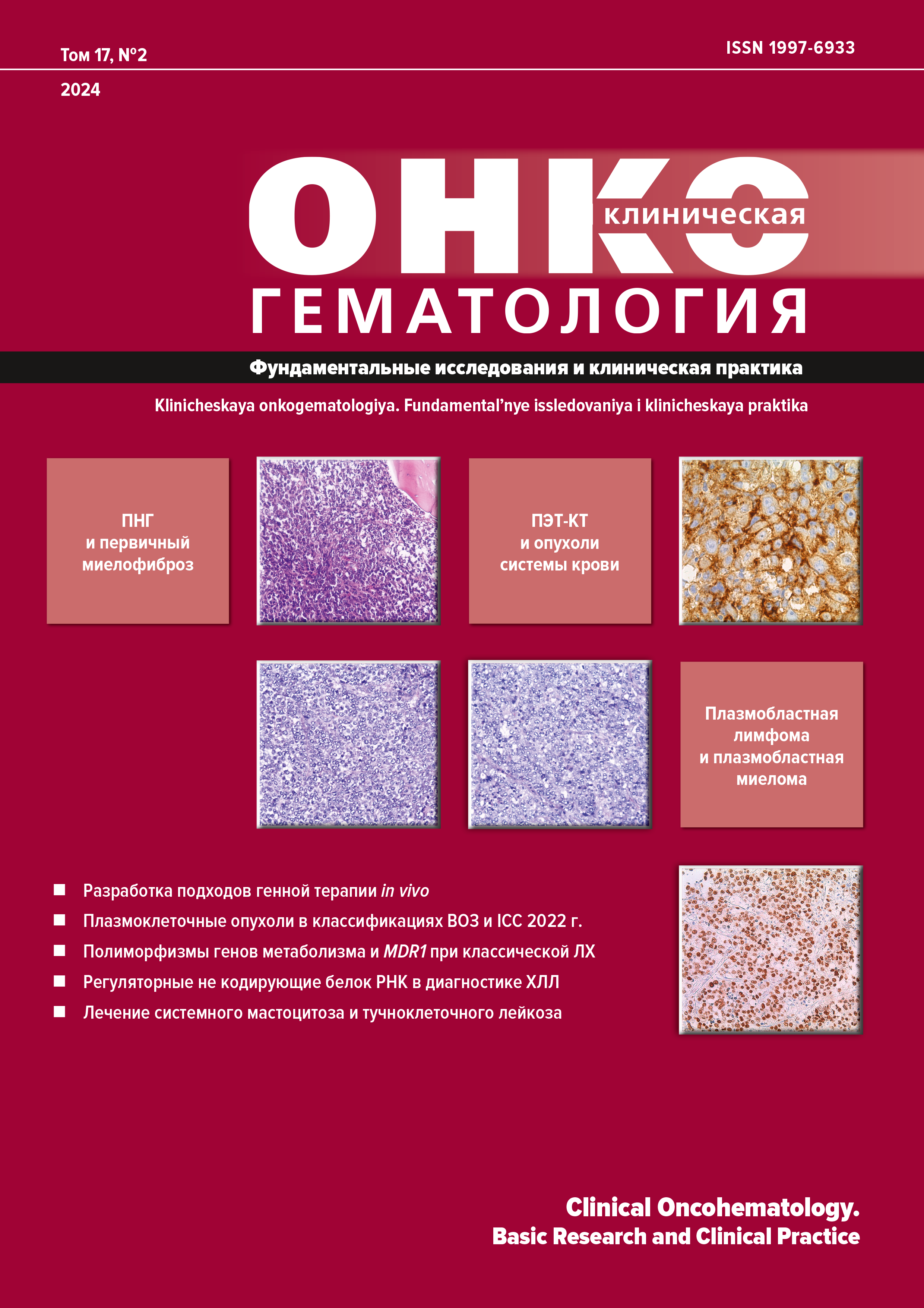
In 2022, the hematological community was faced with a rather non-trivial event of simultaneous publication of two competitive classifications of hematopoietic and lymphoid tumors drawn up by different teams of the international leading experts. During the last 20 years, the generally recognized standard used for diagnosis was provided by several consecutive editions of classifications of hematological neoplasms published by the World Health Organization (WHO) in 2001, 2008, and 2016. Since the 4th edition of the WHO classification (WHO-HAEM4), new clinicopathologic, biological, and molecular knowledge has accumulated in this area, which promoted the refinement of diagnostic criteria for some diseases, the emergence of new terms, and the endorsement of notions previously defined as requiring further clarification. As a result, the next 5th edition of the WHO classification of tumours of haematopoietic and lymphoid tissues (WHO-HAEM5) was prepared and published as a preliminary article in the Leukemia. In this regard, it is worth noting that the final version of the WHO Blue Book was not released in 2023 and, therefore, can still be accomplished by some additions. Furthermore, in the same year of 2022, the Blood published the article “The International Consensus Classification of Mature Lymphoid Neoplasms” abbreviated to ICC. The authors of the two classifications hardly overlap. The present review compares these classifications with regard to new diagnostic criteria and verification of concrete clinicopathologic categories. The review largely focuses on plasma cell tumors and related B-cell lymphoproliferative diseases characterized by monoclonal immunoglobulin secretion.
References
- Alaggio R, Amador C, Anagnostopoulos I, et al. The 5th edition of the World Health Organization Classification of Haematolymphoid Tumours: Lymphoid Neoplasms. Leukemia. 2022;36(7):1720–48. doi: 10.1038/s41375-022-01620-2.
- Campo E, Jaffe ES, Cook JR, et al. The International Consensus Classification of Mature Lymphoid Neoplasms: a report from the Clinical Advisory Committee. Blood. 2022;140(11):1229–53. doi: 10.1182/blood.2022015851.
- Kyle RA, Ansell SM, Kapoor P. Prognostic factors and indications for treatment of Waldenstrom’s Macroglobulinemia. Best Pract Res Clin Haematol. 2016;29(2):179–86. doi: 10.1016/j.beha.2016.08.014.
- Khwaja J, D’Sa S, Minnema MC, et al. IgM monoclonal gammopathies of clinical significance: diagnosis and management. Haematologica. 2022;107(9):2037–50. doi: 10.3324/haematol.2022.280953.
- Annibali O, Petrucci MT, Del Bianco P, et al. IgM multiple myeloma: report of four cases and review of the literature. Leuk Lymphoma. 2006;47(8):1565–9. doi: 10.1080/10428190600604450.
- Kyle RA, Anderson KC. A tribute to Jan Gosta Waldenstrom. Blood. 1997;89(12):4245–7.
- Owen RG, Treon SP, Al-Katib A, et al. Clinicopathological definition of Waldenstrom’s macroglobulinemia: consensus panel recommendations from the Second International Workshop on Waldenstrom’s Macroglobulinemia. Semin Oncol. 2003;30(2):110–5. doi: 10.1053/sonc.2003.50082.
- Fend F, Dogan A, Cook JR. Plasma cell neoplasms and related entities-evolution in diagnosis and classification. Virchows Arch. 2023;482(1):163–77. doi: 10.1007/s00428-022-03431-3.
- Garcia-Abellas P, Ferrer Gomez A, Bueno Sacristan D, et al. Lymphoplasmacytic lymphoma and marginal zone lymphoma involving bone marrow: A diagnostic dilemma. Useful clinicopathological features to accurate the diagnosis. EJHaem. 2022;3(4):1181–7. doi: 10.1002/jha2.573.
- Kivrak H, Yuksel S, Ates C, et al. Relevance of Additional Immunohistochemical Markers in the Differential Diagnosis of Small B-Cell Lymphomas: A Case-Control Study. Turk J Haematol. 2022;39(3):178–87. doi: 10.4274/tjh.galenos.2021.2021.0349.
- Gertz MA. Waldenstrom macroglobulinemia: 2023 update on diagnosis, risk stratification, and management. Am J Hematol. 2023;98(2):348–58. doi: 10.1002/ajh.26796.
- Treon SP, Xu L, Guerrera ML, et al. Genomic Landscape of Waldenstrom Macroglobulinemia and Its Impact on Treatment Strategies. J Clin Oncol. 2020;38(11):1198–208. doi: 10.1200/JCO.19.02314.
- Deguine J, Barton GM. MyD88: a central player in innate immune signaling. F1000Prime Rep. 2014;6:97. doi: 10.12703/P6-97.
- Awata-Shiraiwa M, Yokohama A, Kanai Y, et al. Waldenstrom macroglobulinemia and non-IgM-type lymphoplasmacytic lymphoma are genetically similar. Acta Haematol. 2023;146(5):384–90. doi: 10.1159/000530100.
- Treon SP, Xu L, Hunter Z. MYD88 Mutations and Response to Ibrutinib in Waldenstrom’s Macroglobulinemia. N Engl J Med. 2015;373(6):584–6. doi: 10.1056/NEJMc1506192.
- Kaiser LM, Hunter ZR, Treon SP, Buske C. CXCR4 in Waldenstrom’s Macroglobulinema: chances and challenges. Leukemia. 2021;35(2):333–45. doi: 10.1038/s41375-020-01102-3.
- Alcoceba M, Garcia-Alvarez M, Medina A, et al. MYD88 Mutations: Transforming the Landscape of IgM Monoclonal Gammopathies. Int J Mol Sci. 2022;23(10):5570. doi: 10.3390/ijms23105570.
- Garcia-Sanz R, Dogliotti I, Zaccaria GM, et al. 6q deletion in Waldenstrom macroglobulinaemia negatively affects time to transformation and survival. Br J Haematol. 2021;192(5):843–52. doi: 10.1111/bjh.17028.
- Krzisch D, Guedes N, Boccon-Gibod C, et al. Cytogenetic and molecular abnormalities in Waldenstrom’s macroglobulinemia patients: Correlations and prognostic impact. Am J Hematol. 2021;96(12):1569–79. doi: 10.1002/ajh.26339.
- Nguyen-Khac F, Lambert J, Chapiro E, et al. Chromosomal aberrations and their prognostic value in a series of 174 untreated patients with Waldenstrom’s macroglobulinemia. Haematologica. 2013;98(4):649–54. doi: 10.3324/haematol.2012.070458.
- Hill QA, Stamps R, Massey E, et al. Guidelines on the management of drug-induced immune and secondary autoimmune, haemolytic anaemia. Br J Haematol. 2017;177(2):208–20. doi: 10.1111/bjh.14654.
- Jager U, Barcellini W, Broome CM, et al. Diagnosis and treatment of autoimmune hemolytic anemia in adults: Recommendations from the First International Consensus Meeting. Blood Rev. 2020;41:100648. doi: 10.1016/j.blre.2019.100648.
- Roelcke D. Cold agglutination. Antibodies and antigens. Clin Immunol Immunopathol. 1974;2(2):266–80. doi: 10.1016/0090-1229(74)90044-0.
- Berentsen S, Ulvestad E, Langholm R, et al. Primary chronic cold agglutinin disease: a population based clinical study of 86 patients. Haematologica. 2006;91(4):460–6.
- Berentsen S. How I treat cold agglutinin disease. Blood. 2021;137(10):1295–303. doi: 10.1182/blood.2019003809.
- Randen U, Troen G, Tierens A, al. Primary cold agglutinin-associated lymphoproliferative disease: a B-cell lymphoma of the bone marrow distinct from lymphoplasmacytic lymphoma. Haematologica. 2014;99(3):497–504. doi: 10.3324/haematol.2013.091702.
- Malecka A, Troen G, Tierens A, et al. Immunoglobulin heavy and light chain gene features are correlated with primary cold agglutinin disease onset and activity. Haematologica. 2016;101(9):e361–e364. doi: 10.3324/haematol.2016.146126.
- Berentsen S. New Insights in the Pathogenesis and Therapy of Cold Agglutinin-Mediated Autoimmune Hemolytic Anemia. Front Immunol. 2020;11:590. doi: 10.3389/fimmu.2020.00590.
- Malecka A, Delabie J, Ostlie I, et al. Cold agglutinin-associated B-cell lymphoproliferative disease shows highly recurrent gains of chromosome 3 and 12 or 18. Blood Adv. 2020;4(6):993–6. doi: 10.1182/bloodadvances.2020001608.
- Malecka A, Troen G, Tierens A, et al. Frequent somatic mutations of KMT2D (MLL2) and CARD11 genes in primary cold agglutinin disease. Br J Haematol. 2018;183(5):838–42. doi: 10.1111/bjh.15063.
- Boyle EM, Deshpande S, Tytarenko R, et al. The molecular make up of smoldering myeloma highlights the evolutionary pathways leading to multiple myeloma. Nat Commun. 2021;12(293):2. doi: 10.1038/s41467-41020-20524-41462.
- Rajkumar SV, Dimopoulos MA, Palumbo A, et al. International Myeloma Working Group updated criteria for the diagnosis of multiple myeloma. Lancet Oncol. 2014;15(12):e538–e548. doi: 10.1016/S1470-2045(14)70442-5.
- Kyle RA, Larson DR, Therneau TM, et al. Long-Term Follow-up of Monoclonal Gammopathy of Undetermined Significance. N Engl J Med. 2018;378(3):241–9. doi: 10.1056/NEJMoa1709974.
- Al Hamed R, Bazarbachi AH, Bazarbachi A, et al. Comprehensive Review of AL amyloidosis: some practical recommendations. Blood Cancer J. 2021;11(5):97. doi: 10.1038/s41408-021-00486-4.
- Leung N, Bridoux F, Batuman V, et al. The evaluation of monoclonal gammopathy of renal significance: a consensus report of the International Kidney and Monoclonal Gammopathy Research Group. Nat Rev Nephrol. 2019;15(1):45–59. doi: 10.1038/s41581-018-0077-4.
- Смирнов А.В., Афанасьев Б.В., Поддубная И.В. и др. Моноклональная гаммапатия ренального значения: консенсус гематологов и нефрологов России по введению нозологии, диагностике и обоснованности клон-ориентированной терапии. Нефрология. 2019;23(6):9–28. doi: 10.36485/1561-6274-2019-236-9-28. [Smirnov AV, Afanasyev BV, Poddubnaya IV, et al. Monoclonal gammopathy of renal significance: Consensus of hematologists and nephrologists of Russia on the establishment of nosology, diagnostic approach and rationale for clone specific treatment. Nephrology (Saint-Petersburg). 2019;23(6):9–28. doi: 10.36485/1561-6274-2019-236-9-28. (In Russ)]
- Amaador K, Peeters H, Minnema MC, et al. Monoclonal gammopathy of renal significance (MGRS) histopathologic classification, diagnostic workup, and therapeutic options. Neth J Med. 2019;77(7):243–54.
- Leung N, Bridoux F, Hutchison CA, et al. Monoclonal gammopathy of renal significance: when MGUS is no longer undetermined or insignificant. Blood. 2012;120(22):4292–5. doi: 10.1182/blood-2012-07-445304.
- Rajkumar SV, Kumar S, Lonial S, Mateos MV. Smoldering multiple myeloma current treatment algorithms. Blood Cancer J. 2022;12(9):129. doi: 10.1038/s41408-022-00719-0.
- Ravindran A, Bartley AC, Holton SJ, et al. Prevalence, incidence and survival of smoldering multiple myeloma in the United States. Blood Cancer J. 2016;6(10):e486. doi: 10.1038/bcj.2016.100.
- Kyle RA, Remstein ED, Therneau TM, et al. Clinical course and prognosis of smoldering (asymptomatic) multiple myeloma. N Engl J Med. 2007;356(25):2582–90. doi: 10.1056/NEJMoa070389.
- Lakshman A, Rajkumar SV, Buadi FK, et al. Risk stratification of smoldering multiple myeloma incorporating revised IMWG diagnostic criteria. Blood Cancer J. 2018;8(6):59. doi: 10.1038/s41408-018-0077-4.
- Vaxman I, Gertz MA. How I approach smoldering multiple myeloma. Blood. 2022;140(8):828–38. doi: 10.1182/blood.2021011670.
- Moreau P, San Miguel J, Sonneveld P, et al. Multiple myeloma: ESMO Clinical Practice Guidelines for diagnosis, treatment and follow-up. Ann Oncol. 2017;28(Suppl_4):iv52–iv61. doi: 10.1093/annonc/mdx096.
- Kind S, Merenkow C, Buscheck F, et al. Prevalence of Syndecan-1 (CD138) Expression in Different Kinds of Human Tumors and Normal Tissues. Dis Markers. 2019;2019:4928315. doi: 10.1155/2019/4928315.
- Flores-Montero J, de Tute R, Paiva B, et al. Immunophenotype of normal vs. myeloma plasma cells: Toward antibody panel specifications for MRD detection in multiple myeloma. Cytometry B Clin Cytom. 2016;90(1):61–72. doi: 10.1002/cyto.b.21265.
- An G, Xu Y, Shi L, et al. t(11;14) multiple myeloma: a subtype associated with distinct immunological features, immunophenotypic characteristics but divergent outcome. Leuk Res. 2013;37(10):1251–7. doi: 10.1016/j.leukres.2013.06.020.
- Fonseca R, Bergsagel PL, Drach J, et al. International Myeloma Working Group molecular classification of multiple myeloma: spotlight review. Leukemia. 2009;23(12):2210–21. doi: 10.1038/leu.2009.174.
- Tian E, Sawyer JR, Heuck CJ, et al. In multiple myeloma, 14q32 translocations are nonrandom chromosomal fusions driving high expression levels of the respective partner genes. Genes Chromosomes Cancer. 2014;53(7):549–57. doi: 10.1002/gcc.22165.
- Bal S, Kumar SK, Fonseca R, et al. Multiple myeloma with t(11;14): unique biology and evolving landscape. Am J Cancer Res. 2022;12(7):2950–65.
- Prideaux SM, Conway O’Brien E, Chevassut TJ. The genetic architecture of multiple myeloma. Adv Hematol. 2014;2014:864058. doi: 10.1155/2014/864058.
- Fonseca R, Barlogie B, Bataille R, et al. Genetics and cytogenetics of multiple myeloma: a workshop report. Cancer Res. 2004;64(4):1546–58. doi: 10.1158/0008-5472.can-03-2876.
- Abdallah N, Rajkumar SV, Greipp P, et al. Cytogenetic abnormalities in multiple myeloma: association with disease characteristics and treatment response. Blood Cancer J. 2020;10(8):82. doi: 10.1038/s41408-020-00348-5.
- D’Agostino M, Cairns DA, Lahuerta JJ, et al. Second Revision of the International Staging System (R2-ISS) for Overall Survival in Multiple Myeloma: A European Myeloma Network (EMN) Report Within the HARMONY Project. J Clin Oncol. 2022;40(29):3406–18. doi: 10.1200/JCO.21.02614.
- Войцеховский В.В., Григоренко А.А., Есенина Т.В. и др. Особенности диагностики и лечения различных вариантов плазмоцитомы. Бюллетень физиологии и патологии дыхания. 2023;88:105–19. doi: 10.36604/1998-5029-2023-88-105-119. [Voytsekhovskiy VV, Grigorenko AA, Esenina TV, et al. Features of diagnostics and treatment of various plasmacytoma options. Bulletin Physiology and Pathology of Respiration. 2023;88:105–19. doi: 10.36604/1998-5029-2023-88-105-119. (In Russ)]
- Caers J, Paiva B, Zamagni E, et al. Diagnosis, treatment, and response assessment in solitary plasmacytoma: updated recommendations from a European Expert Panel. J Hematol Oncol. 2018;11(1):10. doi: 10.1186/s13045-017-0549-1.
- Nahi H, Genell A, Walinder G, et al. Incidence, characteristics, and outcome of solitary plasmacytoma and plasma cell leukemia. Population-based data from the Swedish Myeloma Register. Eur J Haematol. 2017;99(3):216–22. doi: 10.1111/ejh.12907.
- Swerdlow SH, Campo E, Pileri SA, et al. The 2016 revision of the World Health Organization classification of lymphoid neoplasms. Blood. 2016;127(20):2375–90. doi: 10.1182/blood-2016-01-643569.
- De Waal EG, Leene M, Veeger N, et al. Progression of a solitary plasmacytoma to multiple myeloma. A population-based registry of the northern Netherlands. Br J Haematol. 2016;175(4):661–7. doi: 10.1111/bjh.14291.
- Kremer M, Ott G, Nathrath M, et al. Primary extramedullary plasmacytoma and multiple myeloma: phenotypic differences revealed by immunohistochemical analysis. J Pathol. 2005;205(1):92–101. doi: 10.1002/path.1680.
- Firsova MV, Mendeleeva LP, Kovrigina AM, et al. Plasmacytoma in patients with multiple myeloma: morphology and immunohistochemistry. BMC Cancer. 2020;20(1):346. doi: 10.1186/s12885-020-06870-w.
- Boll M, Parkins E, O’Connor SJ, et al. Extramedullary plasmacytoma are characterized by a ‘myeloma-like’ immunophenotype and genotype and occult bone marrow involvement. Br J Haematol. 2010;151(5):525–7. doi: 10.1111/j.1365-2141.2010.08386.x.
- Mori H, Fukatsu M, Ohkawara H, et al. Heterogeneity in the diagnosis of plasmablastic lymphoma, plasmablastic myeloma, and plasmablastic neoplasm: a scoping review. Int J Hematol. 2021;114(6):639–52. doi: 10.1007/s12185-021-03211-w.
- Gowin K, Skerget S, Keats JJ, et al. Plasma cell leukemia: A review of the molecular classification, diagnosis, and evidenced-based treatment. Leuk Res. 2021;111:106687. doi: 10.1016/j.leukres.2021.106687.
- Sant M, Allemani C, Tereanu C, et al. Incidence of hematologic malignancies in Europe by morphologic subtype: results of the HAEMACARE project. Blood. 2010;116(19):3724–34. doi: 10.1182/blood-2010-05-282632.
- Gonsalves WI, Rajkumar SV, Go RS, et al. Trends in survival of patients with primary plasma cell leukemia: a population-based analysis. Blood. 2014;124(6):907–12. doi: 10.1182/blood-2014-03-565051.
- Kyle RA, Maldonado JE, Bayrd ED. Plasma cell leukemia. Report on 17 cases. Arch Intern Med. 1974;133(5):813–8. doi: 10.1001/archinte.133.5.813.
- Granell M, Calvo X, Garcia-Guinon A, et al. Prognostic impact of circulating plasma cells in patients with multiple myeloma: implications for plasma cell leukemia definition. Haematologica. 2017;102(6):1099–104. doi: 10.3324/haematol.2016.158303.
- Ravi P, Kumar SK, Roeker L, et al. Revised diagnostic criteria for plasma cell leukemia: results of a Mayo Clinic study with comparison of outcomes to multiple myeloma. Blood Cancer J. 2018;8(12):116. doi: 10.1038/s41408-018-0140-1.
- Fernandez de Larrea C, Kyle R, Rosinol L, et al. Primary plasma cell leukemia: consensus definition by the International Myeloma Working Group according to peripheral blood plasma cell percentage. Blood Cancer J. 2021;11(12):192. doi: 10.1038/s41408-021-00587-0.
- Bardwick PA, Zvaifler NJ, Gill GN, et al. Plasma cell dyscrasia with polyneuropathy, organomegaly, endocrinopathy, M protein, and skin changes: the POEMS syndrome. Report on two cases and a review of the literature. Medicine (Baltimore). 1980;59(4):311–22. doi: 10.1097/00005792-198007000-00006.
- Dispenzieri A. POEMS syndrome: 2021 Update on diagnosis, risk-stratification, and management. Am J Hematol. 2021;96(7):872–88. doi: 10.1002/ajh.26240.
- Пирадов М.А., Супонева Н.А., Гинзберг М.А. и др. POEMS-синдром: обзор литературы и описание клинических наблюдений. Журнал неврологии и психиатрии им. С.С. Корсакова. 2014;114(4):4–10. [Piradov MA, Suponeva NA, Ginzberg MA, et al. POEMS-syndrome: a literature review and case reports. Zhurnal Nevrologii i Psikhiatrii imeni S.S. Korsakova. 2014;114(4):4–10. (In Russ)]
- Михайлов А.М., Бессмельцев С.С., Пожарисский К.М. и др. Болезнь Каслмана и POEMS-синдром. Клиническая онкогематология. 2010;3(3):259–69. [Mihailov AM, Bessmeltcev SS, Pojarisskiy KM, et al. Castleman’s disease and POEMS-syndrome. Klinicheskaya onkogematologiya. 2010;3(3):259–69. (In Russ)]
- Sykes DB, O’Connell C, Schroyens W. The TEMPI syndrome. Blood. 2020;135(15):1199–203. doi: 10.1182/blood.2019004216.
- Lipsker D. Monoclonal gammopathy of cutaneous significance: review of a relevant concept. J Eur Acad Dermatol Venereol. 2017;31(1):45–52. doi: 10.1111/jdv.13847.
- Rosado FG, Oliveira JL, Sohani AR, et al. Bone marrow findings of the newly described TEMPI syndrome: when erythrocytosis and plasma cell dyscrasia coexist. Mod Pathol. 2015;28(3):367–72. doi: 10.1038/modpathol.2014.117.
- Lipsker D, Rondeau M, Massard G, Grosshans E. The AESOP (adenopathy and extensive skin patch overlying a plasmacytoma) syndrome: report of 4 cases of a new syndrome revealing POEMS (polyneuropathy, organomegaly, endocrinopathy, monoclonal protein, and skin changes) syndrome at a curable stage. Medicine (Baltimore). 2003;82(1):51–9. doi: 10.1097/00005792-200301000-00005.
- Lenormand C, Marzolf G, Lipsker D. AESOP syndrome: a potential life-saving and early clue to the diagnosis of POEMS syndrome. Clin Dermatol. 2021;39(2):215–9. doi: 10.1016/j.clindermatol.2020.10.002.
- Dagrosa AT, Cowdrey MCE, LeBlanc RE, et al. Adenopathy and extensive skin patch overlying a plasmacytoma with unusual histologic findings in a patient with polyneuropathy, organomegaly, endocrinopathy, monoclonal protein and skin changes syndrome and Castleman disease. J Cutan Pathol. 2019;46(10):784–9. doi: 10.1111/cup.13514.
- Быкова В.П., Дайхес Н.А., Платонова Г.А. и др. Опухолевидный амилоидоз верхних дыхательных путей. Архив патологии. 2019;81(5):74–9. doi: 10.17116/patol20198105174. [Bykova VP, Daikhes NA, Platonova GA, et al. Tumor-like amyloidosis of the upper respiratory tract. Arkhiv patologii. 2019;81(5):74–9. doi: 10.17116/patol20198105174. (In Russ)]
- Basset M, Hummedah K, Kimmich C, et al. Localized immunoglobulin light chain amyloidosis: Novel insights including prognostic factors for local progression. Am J Hematol. 2020;95(10):1158–69. doi: 10.1002/ajh.25915.
- Охота В.К., Рыжко В.В., Ковригина А.М. и др. Болезнь тяжелых цепей-μ в сочетании с системным амилоидозом и неамилоидными депозитами. Трудности диагностики и терапии. Гематология и трансфузиология. 2020;65(2):190–207. doi: 10.35754/0234-5730-2020-65-2-190-207. [Okhota VK, Ryzhko VV, Kovrigina AM, et al. μ-Heavy chain disease associated with systemic amyloidosis and non-amyloid deposits. Difficulties in diagnosis and therapy. Russian journal of hematology and transfusiology. 2020;65(2):190–207. doi: 10.35754/0234-5730-2020-65-2-190-207. (In Russ)]

This work is licensed under a Creative Commons Attribution-NonCommercial-ShareAlike 4.0 International License.
Copyright (c) 2024 Clinical Oncohematology