Keywords
nodular sclerosis
reactive tumor microenvironment
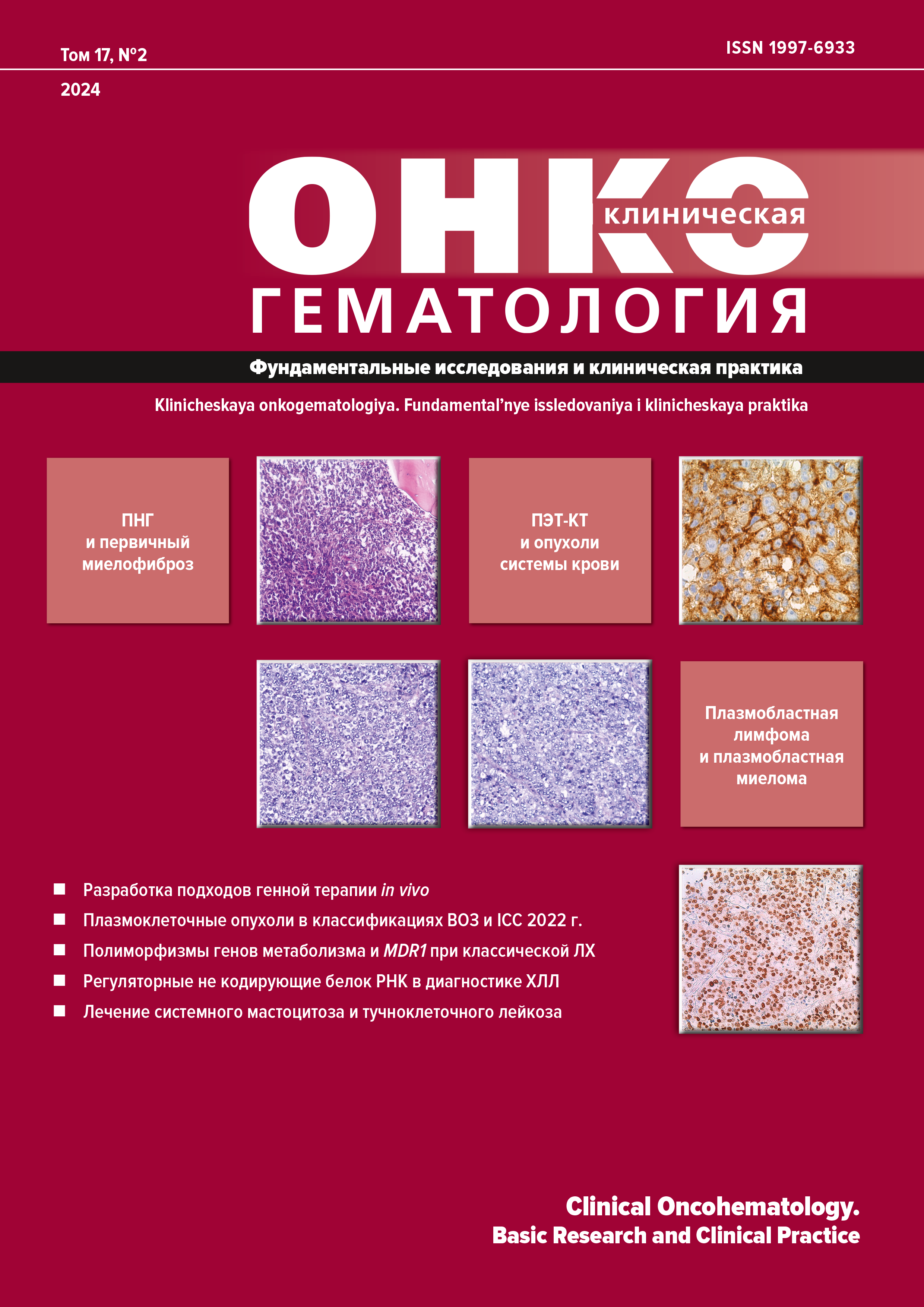
CD20
В-lymphocytes
prognosis
How to Cite
Statistics
PDF_2024-17-2_166-172 (Russian) downloads: 180
Keywords
Abstract
Aim. To assess the prognostic value of CD20-positive B-lymphocytes in the reactive tumor microenvironment using histological specimens of lymph nodes from patients with nodular sclerosis classical Hodgkin lymphoma (cHL).
Materials & Methods. The analysis focused on archival samples (paraffin blocks and cassettes) of lymph nodes from 71 patients with newly diagnosed cHL, nodular sclerosis type. Immunohistochemical methods were used for morphometric assessment of CD20-positive cells in the tumor microenvironment. In each lymph node biopsy sample, 20 fields of view were analyzed for the mean relative count of CD20-positive B-lymphocytes in the reactive tumor microenvironment. The В-cell count was determined by the double-blind method. Patients were aged 18–77 years (median 35 years); there were 37 women and 34 men. According to the primary documentation, 11 (15.5 %) patients received ABVD as first-line therapy and 60 (84.5 %) patients were treated with BEACOPP-14 (EACOPP-14) or BEACOPPesc regimens.
Results. A low count of CD20-positive B-lymphocytes in the reactive tumor microenvironment is regarded as an independent factor of poor prognosis in patients with nodular sclerosis cHL. This is the category of patients who show worse event-free survival (EFS) rates. In the cohort with the subthreshold count of CD20-positive B-lymphocytes in the reactive microenvironment, the median EFS was 38 months, whereas in the cohort with the above-threshold count it was not reached.
Conclusion. The results of this study demonstrate the need to assess the relative count of CD20-positive B-lymphocytes in the reactive tumor microenvironment in patients with nodular sclerosis cHL at the primary diagnosis stage. There is every reason to believe that for this category of patients the incorporation of this immunohistochemical parameter into the existing predictive models is fully justified.
References
- Scott D, Steidl C. The classical Hodgkin lymphoma tumor microenvironment: macrophages and gene expression-based modeling. Hematology Am Soc Hematol Educ Program. 2014;2014(1):144–50. doi: 10.1182/asheducation-2014.1.144.
- Menendez V, Solorzano JL, Fernandez S, et al. The Hodgkin Lymphoma Immune Microenvironment: Turning Bad News into Good. Cancers (Basel). 2022;5(14):1360. doi: 10.3390/cancers14051360.
- Перфилова Е.А., Минаев М.С., Дьяконов Д.А. и др. Прогностическое значение экспрессии CD163- и CD68-положительных макрофагов при нодулярном склерозе лимфомы Ходжкина. Онкогематология. 2022;17(1):104–12. doi: 10.17650/1818-8346-2022-17-1-104-112. [Perfilova EA, Minaev MS, Diakonov DA, et al. Prognostic significance of CD163- and CD68-expressing macrophages in nodular sclerosis Hodgkin lymphoma. Oncohematology. 2022;17(1):104–12. doi: 10.17650/1818-8346-2022-17-1-104-112. (In Russ)]
- Connors JM, Cozen W, Steidl C, et al. Hodgkin Lymphoma. Nat Rev Dis Primers. 2020;6(1):61. doi: 10.1038/s41572-020-0203-z.
- Swerdlow SH, Campo E, Harris NL, et al. WHO Classification of Tumours of Haematopoietic and Lymphoid Tissues. Revised 4th edition. Lyon: IARC Press; 2017. 586 p.
- Злокачественные новообразования в России в 2020 году (заболеваемость и смертность). Под ред. А.Д. Каприна, В.В. Старинского, Г.В. Петровой. М.: МНИОИ им. П.А. Герцена — филиал ФГБУ «НМИЦ радиологии» Минздрава России, 2021. 252 с. [Kaprin AD, Starinskii VV, Petrova GV, eds. Zlokachestvennye novoobrazovaniya v Rossii v 2020 godu (zabolevaemost’ i smertnost’). (Malignant neoplasms in Russia in 2020 (incidence and mortality.) Moscow: MNIOI im. P.A. Gertsena — filial FGBU “NMITs radiologii” Publ.; 2021. 252 р. (In Russ)]
- Ferhanoglu B, Kim T, Karduss A, et al. Treatment pathways and clinical outcomes in Hodgkin lymphoma outside Europe and North America: results from the international, multicenter, retrospective, B-HOLISTIC study. Leuk Lymphoma. 2022;63(14):3317–30. doi: 10.1080/10428194.2022.2126281.
- Pileri S, Fiori S, Iuliis V, Tabanelli V. Lymphoma, Hodgkin’s: Pathology and Genetics. In: Reference Module in Biomedical Sciences. Elsevier; 2019. doi: 10.1016/B978-0-12-801238-3.65258-3.
- Van Spronsen DJ, Vrints LW, Hofstra G, et al. Disappearance of prognostic significance of histopathological grading of nodular sclerosing Hodgkin’s disease for unselected patients, 1972–92. Br J Haematol. 1997;96(2):322–7. doi: 10.1046/j.1365-2141.1997.d01-2010.x.
- Hess JL, Bodis S, Pinkus G, et al. Histopathologic grading of nodular sclerosis Hodgkin’s disease. Lack of prognostic significance in 254 surgically staged patients. Cancer. 1994;74(2):708–14. doi: 10.1002/1097-0142(19940715)74:2<708::aid-cncr2820740226>3.0.co;2-7.
- Shang J, Zha H, Sun Y. Phenotypes, Functions, and Clinical Relevance of Regulatory B Cells in Cancer. Front Immunol. 2020;11:582657. doi: 10.3389/fimmu.2020.582657.
- Downs-Canner SM, Meier J, Vincent BG, et al. B Cell Function in the Tumor Microenvironment. Annu Rev Immunol. 2022;40:169–93. doi: 10.1146/annurev-immunol-101220-015603.
- Santos M, Lima MM. CD20 role in pathophysiology of Hodgkin’s disease. Rev Assoc Med Bras. 2017;63(9):810–3. doi: 10.1590/1806-9282.63.09.810.
- Vardhana S, Younes A. The immune microenvironment in Hodgkin lymphoma: T cells, B cells, and immune checkpoints. Haematologica. 2016;101(7):794–802. doi: 10.3324/haematol.2015.132761.
- Panico L, Tenneriello V, Ronconi F, et al. High CD20+ background cells predict a favorable outcome in classical Hodgkin lymphoma and antagonize CD68+ macrophages. Leuk Lymphoma. 2015;6(56):1636–42. doi: 10.3109/10428194.2014.951849.
- Greaves P, Clear A, Coutinho R, et al. Expression of FOXP3, CD68, and CD20 at diagnosis in the microenvironment of classical Hodgkin lymphoma is predictive of outcome. J Clin Oncol. 2017;35(10):1140. doi: 10.1200/JCO.2011.39.9881.
- Jachimowicz RD, Pieper L, Reinke S, et al. Whole-slide image analysis of the tumor microenvironment identifies low B-cell content as a predictor of adverse outcome in patients with advanced-stage classical Hodgkin lymphoma treated with BEACOPP. Haematologica. 2021;106(6):1684–92. doi: 10.3324/haematol.2019.243287.
- Jones RJ, Gocke CD, Kasamon YL, et al. Circulating clonotypic B cells in classic Hodgkin lymphoma. Blood. 2009;113(23):5920. doi: 10.1182/blood-2008-11-189688.
- Zhao X, Ma Y, Bian H, et al. CD20 expression is closely associated with Epstein-Barr virus infection and an inferior survival in nodular sclerosis classical Hodgkin lymphoma. Front Oncol. 2022;12:993768. doi: 10.3389/fonc.2022.993768.
- Bertuzzi C, Sabattini E, Agostinelli C. Immune Microenvironment Features and Dynamics in Hodgkin Lymphoma. Cancers. 2021;13(14):3634. doi: 10.3390/cancers13143634.
- Lacet DFR, Oliveira CC. The role of immunohistochemistry in the assessment of classical Hodgkin lymphoma microenvironment. Int J Clin Exper Pathol. 2022;10(15):412–24.

This work is licensed under a Creative Commons Attribution-NonCommercial-ShareAlike 4.0 International License.
Copyright (c) 2024 Clinical Oncohematology